
Translate this page into:
Primary empty sella syndrome presenting as hyponatremia
*Corresponding author: Nidhesh Khemchandani, Department of General Medicine, Himalayan Institute of Medical Sciences, Dehradun, Uttarakhand, India. nidheshkhem@gmail.com
-
Received: ,
Accepted: ,
How to cite this article: Khemchandani N. Primary empty sella syndrome presenting as hyponatremia. Indian J Med Sci 2023;75:82-4.
Abstract
Empty sella is often an incidental magnetic resonance imaging (MRI) finding. It may be partial or complete and can be primary or secondary due to intracranial hypertension, radiation exposure or pituitary apoplexy. Most of the patients are asymptomatic but features of panhypopituitarism can develop in some. This is a case report of a 70-year-old female who presented with complaints of vomiting, altered sensorium with irritability, generalized weakness, difficulty in speaking, and one episode of seizure. Blood investigations revealed hyponatremia that was euvolemic. On further evaluation, she was found to have decreased cortisol, decreased follicle stimulating hormone, decreased thyroxine, normal prolactin, and thyroid-stimulating hormone values suggestive of panhypopituitarism. MRI brain showed an empty sella. Her serum sodium levels improved after starting glucocoticoids and thyroxine tablets, thereby confirming the diagnosis of panhypopituitarism. In the absence of any history of irradiation, hemorrhage, and surgery, a diagnosis of primary empty sella syndrome was made.
Keywords
Empty sella
Persistent hyponatremia
Panhypopituitarism

INTRODUCTION
Empty sella syndrome may be complete or partial depending on whether the sella turcica is completely or partially filled with cerebrospinal fluid. It has certain distinct anatomical and radiological characteristics.[1] Endocrine dysfunction is seen in 25–30% of cases.[2]
In partial empty sella syndrome, pituitary gland thickness ranges from 3 mm to 7 mm and <50% of the sella is filled with cerebrospinal fluid. While in total empty sella syndrome, pituitary gland thickness is <2 mm and more than 50% of the sella is filled with cerebrospinal fluid.[3]
Primary empty sella syndrome is seen commonly in obese and multiparous females and occurs due to combination of increased cerebrospinal fluid pressure with defect in diaphragm sellae.[4]
Empty sella leads to secondary adrenal insufficiency. Adrenal insufficiency can impair water excretion resulting in euvolemic hyponatremia by antidiuretic hormone (ADH) dependent and ADH independent mechanisms. Adrenal insufficiency is associated with elevated ADH levels resulting in hyponatremia.[5]
CASE REPORT
A 70-year-old woman, known case of hypertension (taking Telmisartan) and chronic obstructive pulmonary disease (using metered dose inhaler of Ipratropium and Levosalbutamol), presented to the emergency department with weakness and difficulty in speaking for 4 days along with one episode of vomiting and seizure and altered sensorium with irritability since then. The weakness was generalized, with no involvement of bladder or bowel functions. There was no history of fever. There was no history of any cardiac illness. Her history showed chronic hyponatremia and similar symptoms that improved with intake of sodium-rich diet and administration of normal saline. There was no history of use of diuretics or any pituitary surgery or irradiation. She had achieved menopause at age of 47 years and had two children. Her both deliveries and postpartum periods were uneventful.
On examination, she was irritable but obeying verbal commands, pulse rate was 80/min and blood pressure 160/80 mm Hg. She was afebrile and mildly dehydrated with oxygen saturation of 98% on room air. Jugular venous pressure was normal indicating normal extracellular volume. Neurological examination revealed power of 4/5 in all four limbs with normal deep tendon jerks and flexor plantar response.
Laboratory investigations [Table 1] showed serum creatinine 0.76 mg/dL, serum potassium 3.81 mmol/L, serum sodium 109.75 mmol/L, urinary sodium 86.57 mmol/L, serum osmolality 236.81 mOsmol/kg, and urine osmolality 246.16 mOsmol/kg. Chest X-ray, non-contrast computed tomography head, and abdominal ultrasound were normal. Electrocardiogram was suggestive of left ventricle hypertrophy. Differential diagnosis of syndrome of inappropriate antidiuretic hormone secretion (SIADH) was kept as she had euvolemic hyponatremia. Hypertonic saline was administered but serum sodium levels increased only to 114.56 mmol/L. Restriction of fluids other than hypertonic saline was done along with administration of high-sodium diet. Tolvaptan was initiated at a dose of 15 mg once daily but her serum sodium levels increased only to 120 mmol/L. Endocrine function was investigated again in detail, showing serum follicle stimulating hormone 3.03 mIU/mL, serum prolactin 5.10 ng/ mL, serum free T4 0.6 ng/dL, serum thyroid-stimulating hormone 0.5 microIU/L, and serum cortisol 23.27 ng/ mL suggestive of panhypopituitarism. Magnetic resonance imaging (MRI) brain showed age-related cerebral atrophy with enlarged sella showing cerebrospinal fluid intensity area on all sequences suggestive of empty sella [Figures 1-3]. Her hyponatremia and other symptoms started resolving after administration of Hydrocortisone 100 mg intravenously and Prednisolone 5 mg orally in morning and 2.5 mg orally in evening. Hydrocortisone was given intravenously for 2 days and then stopped. Eltroxin 75 microgram was given once a day orally. The response assessed by her general condition and serum sodium levels was adequate. She was discharged with serum sodium level of 134.04 mmol/L on the above treatment and was asked to come for regular follow-up.

- Magnetic resonance imaging brain T2-weighted image showing empty sella in sagittal section.

- Magnetic resonance imaging brain T1-weighted image showing empty sella in sagittal section.
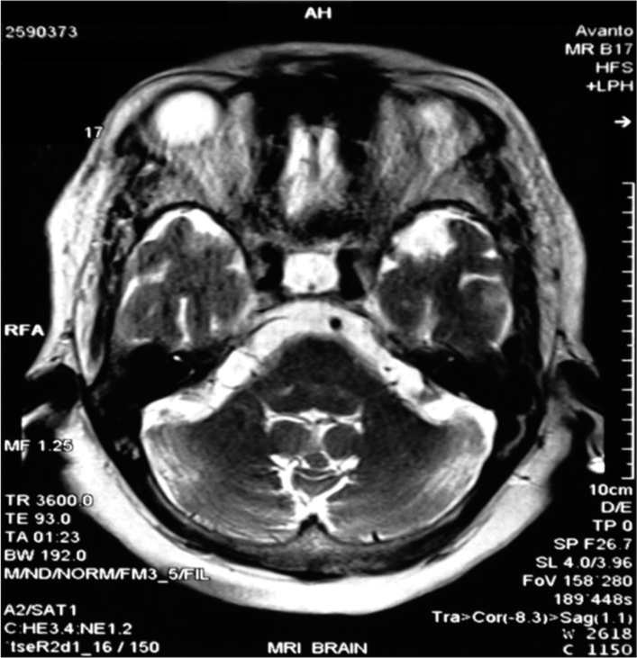
- Magnetic resonance imaging brain showing empty sella with pituitary stalk in axial section.
| Parameter | Patient’s value | Unit |
|---|---|---|
| Hemoglobin | 13.3 | g/dL |
| Total leucocyte count | 15.86 | Thousand/cu mm |
| Plasma glucose | 156 | mg/dL |
| Serum osmolality | 236.81 | mOsmol/kg |
| Serum sodium | 109.75 | mmol/L |
| Serum potassium | 3.8 | mmol/L |
| Blood urea nitrogen | 15.6 | mg/dL |
| Serum creatinine | 0.76 | mg/dl |
| Serum fT4 | 0.57 | ng/dL |
| Serum fT3 | 1.25 | pg/mL |
| Serum TSH | 0.5 | microIU/L |
| Serum FSH | 3.03 | mIU/L |
| Serum cortisol | 23.27 | ng/mL |
| Serum prolactin | 5.10 | ng/mL |
| Urine sodium | 86.57 | mmol/L |
| Urine osmolality | 246.16 | mOsmol/kg |
TSH: Thyroid-stimulating hormone, FSH: Follicle-stimulating hormone
DISCUSSION
Primary empty sella syndrome is an anatomical condition characterized by remodeling of sella turcica and flattening of the pituitary gland resulting from subarachnoid space extension into an intrasellar position. Primary empty sella syndrome is mostly seen in obese, multiparous women.[1]
Clinical features suggest that the pituitary hypofunction associated with the empty sella syndrome in our patient was partial as she managed to live till 70 years of age without hormonal supplements. Hyperprolactinemia and diabetes insipidus may occur in primary empty sella syndrome if the pituitary stalk is compressed by extensive arachnoid herniation. In our patient, there was no compression of pituitary stalk as was evident by normal prolactin levels.
All possible causes of seizures in our patient were ruled out by neurological examination and brain imaging. Neurological examination showed no focal neurological deficit so possibility of any space occupying lesion or cerebrovascular accident was ruled out. Absence of features like history of any head injury or abrupt withdrawal of anti-epileptic drugs, history of fever, history of exposure to any provocative stimuli, and exposure to any toxin or poison with a normal sleep cycle suggested that the possibility of hyponatremia was kept as the precipitating cause for seizures in the patient.
All possible causes of hyponatremia were considered. Based on urinary sodium and urine osmolality reports along with serum osmolality values, SIADH was kept as a differential diagnosis. The response to tolvaptan was not adequate. Despite starting tolvaptan, patient’s serum sodium did not normalize and hyponatremia persisted. Hence, other causes of euvolemic hyponatremia were looked for and thyroid function tests and serum cortisol levels were done. Thyroid profile showed central hypothyroidism. A detailed endocrinological workup was done which showed decreased serum cortisol and serum follicle-stimulating hormone. A diagnosis of pan hypopituitarism was made and so MRI brain was done. MRI brain showed an empty sella. As there was no history of any prior radiation, pituitary surgery, or post-partum event, a diagnosis of primary empty sella as the cause of hyponatremia due to panhypopituitarism was made.
CONCLUSION
The management of hyponatremia should not only include correction of serum sodium concentration but also an attempt must be made to find out the cause of hyponatremia.
Hypopituitarism should be kept as a differential diagnosis in all cases with SIADH like clinical picture. A proper history and physical examination must be done to find out the cause of hyponatremia.
If central hypothyroidism or adrenal insufficiency is seen, hypopituitarism must be suspected and brain imaging should be done to assess the pituitary gland.
Declaration of patient consent
Patient’s consent not required as patient’s identity is not disclosed or compromised.
Conflicts of interest
There are no conflicts of interest.
Financial support and sponsorship
Nil.
References
- The empty sella: Results of treatment in 76 successive cases and high frequency of endocrine and neurological disturbances. Clin Endocrinol (Oxf). 1992;37:529-33.
- [CrossRef] [PubMed] [Google Scholar]
- Empty sella syndrome-beyond being an incidental finding. Indian J Endocrinol Metab. 2012;16(Suppl 2):S321-3.
- [CrossRef] [PubMed] [Google Scholar]
- Primary empty sella (PES): A review of 175 cases. Pituitary. 2013;16:270-4.
- [CrossRef] [PubMed] [Google Scholar]
- Diagnosis and management of hyponatraemia in hospitalised patients. Int J Clin Pract. 2009;63:1494-508.
- [CrossRef] [PubMed] [Google Scholar]


